Adenovirus 1 in Dogs
Infectious Canine Hepatitis in Dogs
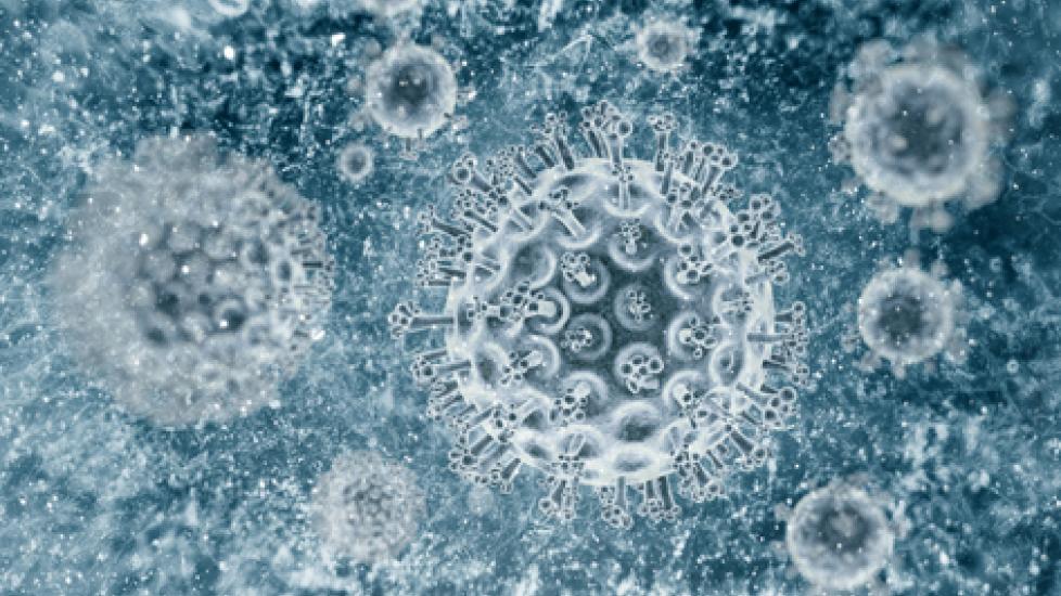
Infectious canine hepatitis is a viral disease of that is caused by the canine adenovirus CAV-1, a type of DNA virus that causes upper respiratory tract infections. This virus targets the parenchymal (functional) parts of the organs, notably the liver, kidneys, eyes and endothelial cells (the cells that line the interior surface of the blood vessels).
The virus begins by localizing in the tonsils around 4 to 8 days after nose and mouth exposure. It then spreads into the bloodstream -- a condition know as viremia (in the blood stream) -- and localizes in the Kupffer cells (specialized white blood cells located in the liver) and endothelium of the liver. Ideally, these white cells, called macrophages, defend the body against infectious invaders, but some viruses have the ability to macropahages as vehicles for replication and spread. CAV-1 is one such virus, taking advantage of the Kupffer cells to replicate and spread, in the process damaging the adjacent hepatocytes (liver cells that are involved in protein synthesis and storage, and transformation of carbohydrates). During this stage of the infection, the virus is shed into the feces and saliva, making both infectious to other dogs.
In a healthy dog with an adequate antibody response, the viral cells will clear the organs in 10 to 14 days, but will remain localized in the kidneys, where the virus will continue to be shed in the urine for 6 to 9 months.
In dogs with only partial neutralizing antibody response, chronic hepatitis takes place. This severe condition often results in cytotoxic ocular injury due to inflammation and death of the cells in the eye with inflammation of the front of the eye (anterior uveitis). This condition leads to one of the more outwardly visible and classic signs of infectious hepatitis: “hepatitis blue eye.”
There are no breed, genetic, or gender associations for acquiring the CAV-1 virus, but but it is primarily seen in dogs that are less than one year of age.
Symptoms
Symptoms will depend on the immunologic status of the host and degree of initial injury to the cells (cytotoxic):
- Peracute (very severe) stage will have symptoms of fever, central nervous system signs, collapse of blood vessels, coagulation disorder (DIC); death frequently occurs within hours
- Acute (severe) stage will show symptoms of fever, anorexia, lethargy, vomiting, diarrhea, enlarged liver, abdominal pain, abdominal fluid, inflammation of the vessels (vasculitis), pinpoint red dots, bruising of skin (petechia), DIC, swollen, enlarged lymph nodes (lymphadenopathy), and rarely, inflammation of the brain (nonsuppurative encephalitis)
- Uncomplicated infection will have symptoms of lethargy, anorexia, transient fever, tonsillitis, vomiting, diarrhea, lymphadenopathy, enlarged liver, abdominal pain
- Late stage infection will result in 20 percent of cases developing eye inflammation and corneal swelling four to six days postinfection; recovery often within 21 days, but may progress to glaucoma and corneal ulceration
Causes
- Contact with infectious CAV-1 adenovirus
- Unvaccinated dogs are at highest risk
Diagnosis
You will need to give a thorough history of your dog's health, onset of symptoms, previous illnesses, and possible incidents that might have led to this condition. Contact with other dogs, such as in kennels, or frequency of contact with feces, such as in open spaces where dogs are permitted to defecate, may play a role in acquiring this virus.
Your veterinarian will perform a thorough physical exam on your dog, with standard laboratory work. A complete blood profile will be conducted, including a chemical blood profile, a complete blood count, a urinalysis and an electrolyte panel. Other laboratory work that will need to be done to confirm a diagnosis of infectious hepatitis include coagulation tests to check for the clotting function of the blood, serology for antibodies to CAV-1, viral isolation of the virus cells, and viral culture. Your doctor will be checking for other common diseases as well, including parvovirus and distemper.
Imaging techniques will include an abdominal radiography to look for enlargement of the liver (hepatomegaly) and fluid buildup in the abdominal cavity, and abdominal ultrasonography, which can give a more detailed view of the liver and whether it is enlarged of is suffering from necrosis (cell death). The latter technique is especially necessary if there is abdominal swelling, as the radiography will show a reduced image detail if there is fluid blocking the view to the liver, where ultrasound imaging will return information based on the depth of frequency of the echo, based on the structure of the tissues. That is, cellular/tissue death in the liver will show decreased echo (hypoechoeic), and severe fluid build up in the abdomen will not return any echoes (anechoic).
A liver biopsy may also need to be performed to make a conclusive diagnosis.
Treatment
If the infection is in the very early stage and is uncomplicated, treatment may be given on an outpatient basis. However, treatment is usually given inpatient. Fluid therapy will be given for electrolyte imbalances that result from vomiting and diarrhea. Potassium and magnesium are often very low and need to be supplement immediately. Blood component therapy will be given for coagulopathy (disorders in the blood's ability to clot). With overt DIC, fresh blood products and low molecular weight heparin will need to be sued to stabilize your dog's condition.
Nutritional support will include giving frequent small meals as tolerated, optimizing nitrogen intake, and feeding the dog according to protein needs. The amount of protein will depend entirely on your dog's individual condition, as some dogs will have high protein in the body and some will have low. Inappropriate protein restriction may impair tissue repair and regeneration. Nitrogen will be restricted if your dog is showing obvious signs of hepatic encephalopathy (a neuropsychiatric abnormality that causes inflammation of the brain and is related to liver failure).
Partial intravenous nutrition will be given for a maximum of five days, or preferably, total intravenous nutrition if oral feeding is not tolerated by the dog. Your doctor will prescribe antibiotics and/or fluid reducers as necessary.
Living and Management
The veterinarian will schedule follow up visits to monitor fluid, electrolyte, acid-base, and coagulation status, and to adjust supportive measures. Sudden kidney failure will also need to be monitored for. A highly digestible diet will need to be fed to your dog during recovery, and a safe place set aside to rest and recover from the illness. Restrict your dog's activity during the recovery period, as well as access to other pets. be especially mindful about cleaning up after your dog, as the virus can continue to be shed long after the recovery period.
Prevention of this infection requires a a modified live virus vaccination for this disease at six to eight weeks of age. The initial vaccination is followed by two booster shots given at three to four weeks apart until the dog reaches 16 weeks of age, with an additional booster given at one year. This is a highly effective vaccine.
Help us make PetMD better
Was this article helpful?
