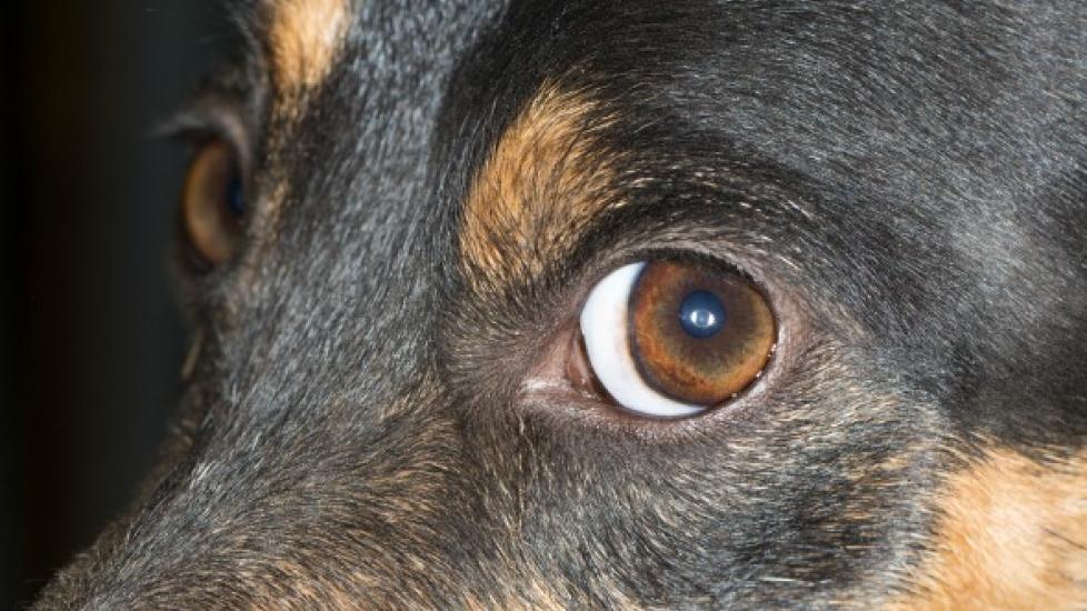
Degeneration of the Cornea in Dogs
Corneal Degenerations and Infiltrations in Dogs
The cornea is the transparent lining that covers the external front of the eyeball; that is, the iris and the pupil (respectively, the colored area that expands and contracts to allow light in, and the lens that transmits the light and image to the brain – the black center). The cornea is continuous with the white part of the eye, the sclera, which covers the rest of the eyeball. Beneath the cornea and the sclera is a layer of connective tissue that supports the eyeball from inside, called the stroma.
Corneal degeneration is a one-sided or two-sided condition, secondary to other eye (ocular) or body (systemic) disorders. It is characterized by lipid (fat-soluble molecules) or calcium deposits within the corneal stroma, and/or epithelium (tissue composed of layers of cells that line the inner hollow of the eyeball, beneath the stroma).
The condition or disease described in this medical article can affect both dogs and cats. If you would like to learn more about how this disease affects cats, please visit this page in the PetMD health library.
Symptoms and Types
The cornea appears rough, with distinct margins where the edge of the cornea meets with the sclera. Associated ocular conditions, such as corneal scars, inflammation of the cornea, or chronic uveitis (long-standing inflammatory disease of the front of the eye), can lead to degeneration of the cornea. If one or more of these conditions have been present, having the cornea checked for further damage would be wise for the prevention of severe and permanent damage.
Causes
One of the main causes of corneal degeneration is lipid (fat) deposits in the supporting structure of the inner eyeball: the stroma and the epithelium. While lipids are a normal part of the body, being, as they are, a principal structure of living cells, hyper deposits of lipids in the tissues can bring about disorders to the system they are inhabiting. Systemic hyperlipoproteinemia, a metabolic disorder characterized by elevated concentrations of cholesterol and specific lipoprotein particles in the blood plasma, may increase the risk of deposits in the stroma, or may worsen already existing deposits. Hyperlipoproteinemia can be secondary to hypothyroidism, diabetes mellitus, hyperadrenocorticism (chronic production of too much cortisone), pancreatitis, nephrotic syndrome (a disorder in which the kidneys are damaged), and liver disease.
Hypercalcemia, a condition that is characterized by the production of too much calcium, can increase the risk of deposits of calcium in the stroma, which can also lead to corneal degeneration. Calcium deposits in the stroma are seen less frequently than lipid deposits.
Other disorders that can affect the cornea and its functionality are hypophosphatemia, an electrolyte irregularity distinguished by too little phosphorus in the blood, and hypervitaminosis D, the production of excess vitamin D.
Corneal degeneration is non-inherited, but it does have a higher rate of incidence with miniature schnauzers.
Diagnosis
Your veterinarian will look for several indications before settling on a diagnosis. Yourdog 's eyes will be coated with fluorescein stain, an orange dye that is viewed in blue light to detect damage to the cornea, or to the presence of foreign objects on the surface of the eye. The stain examination may show a corneal ulcer with varying degrees of edema (swelling). The edema, if present, will appear bluish to gray and can vary in size depending on severity, with indistinct margins. The stain would also show the presence of a corneal scar – which would cause some opacity, appearing gray to white depending on severity. Corneal ulceration may be associated with worsening of the disease, and vision may be affected if the disease is in an advanced state. Severe vision impairment may occur if a primary eye disease, such as uveitis, is found to be present.
If the fluorescein stain does not show any abnormalities, the veterinarian will look for corneal stromal weakness (dystrophy), which affects both eyes, often affecting proportional focus. The cornea will be gray to white in appearance, with distinct margins. This disorder does not retain fluorescein stain and is not associated with inflammation of the eye. If an object has entered the eye (inflammatory cell infiltrate) it will cause the cornea to appear gray to white, with indistinct margins; microscopic examination of the corneal cells will reveal white blood cells, the cells that are responsible for defending the body against foreign materials and infection, indicating that organisms are present in the eye.
Treatment
If a disease of the eye is present, your veterinarian will treat the condition accordingly. Lipid and calcium deposits that impair vision or create discomfort to the eye, either from a roughened surface, or from disruption and ulceration of the corneal epithelium, may benefit from a vigorous corneal scraping, or a superficial removal of part of the cornea (keratectomy). These procedures would be followed by medical management, since deposits are likely to recur in patients following superficial keratectomy surgery. Your dog's diet will also be a consideration. If hyperlipoproteinemia is diagnosed, a low-fat diet would be beneficial for hindering further progression. Your veterinarian will advise you on this. Both methods of treatment can be useful for slowing or stopping progression of the disease.
Living and Management
Your doctor will want to monitor your dog's serum cholesterol and triglycerides to assess the effectiveness of dietary management, if that has been recommended as a maintenance strategy. If a primary disease if present, its will be monitored for progression or regression, and treated according to the indications and comfort needs of your dog.
Help us make PetMD better
Was this article helpful?
