Anisocoria in Dogs
What Is Anisocoria in Dogs?
Anisocoria is a condition in which a dog’s pupils are different sizes. The pupil, a small dark circle at the center of the eye, can adjust its size to let light into the back of the eye to assist in vision.
In low-light conditions, a normal pupil will dilate (get larger) to let in more light, enabling your dog to perceive shadows and get around in dim surroundings. Conversely, in bright sunlight, the pupil will constrict (get smaller), reducing the amount of light hitting the retina so that your dog is able to see clearly.
Normal, healthy pupils are symmetrical and, whether dilating or constricting, they change size simultaneously. When one pupil is notably larger than the other, it is called anisocoria.
The pupil is surrounded by the iris, the colored ring of the eye. It’s the muscles of the iris and the nerves that run through the eye, face, and brain that control how big the pupils are. Several diseases and conditions can impact these nerves and muscles, affecting the ability to constrict and dilate the pupils appropriately. There are also a few conditions that can affect the brain, disrupting the necessary signals required for normal pupil constriction.
While many of these diseases tend to be chronic, if you notice anisocoria occurring suddenly, seek veterinary care as soon as possible. Anisocoria is relatively rare. While it can be harmless, that is not always the case. Sudden onset of anisocoria is considered a medical emergency, especially if it occurs following trauma.
Why Do Dogs Have Different-Sized Pupils in Anisocoria?
When a disease or trauma disrupts the nerves or muscles controlling pupil size, one pupil can become abnormally dilated or constricted. While lighting plays a role in changing pupil sizes, the sympathetic and parasympathetic nervous systems are also involved.
The sympathetic nervous system is often referred to as “fight or flight.” It’s the segment of the animal’s nervous system that gets triggered when faced with danger or intense physical activity. When the sympathetic nervous system is activated, the pupils dilate to allow more light into the retinas, enhancing vision. This is why frightened dogs often have dilated pupils. On the other hand, the parasympathetic nervous system is nicknamed the “rest and digest” response. It gets activated during periods of relaxation. When a dog is eating or resting, the parasympathetic nervous system comes into play, causing the pupils to become smaller.
The mechanisms engaged in changing pupil size entail the collaboration of multiple elements: nerves, muscles, the brain, and the retina. If any of the processes in this pathway are disrupted, abnormalities can occur. These abnormalities might manifest on the eye’s surface, within the iris muscles, along the nerves connected to the eye, within the retina, or even within the brain itself.
Symptoms Associated with Anisocoria in Dogs
Anisocoria means that the pupils are unequal sizes, signifying that one pupil is either abnormally dilated or constricted.
There are several clinical signs that can occur with anisocoria in dogs, depending on the underlying cause. In addition to asymmetrical pupil sizes, one or many of the following may be noticed:
-
Redness of the conjunctiva, the white part of the eyeball
-
Squinting
-
Drainage from the eye
-
Eye discoloration
-
Drooping of the eyelid or face
-
Head shaking
-
Elevated third eyelid
-
Lethargy or reduced activity level
Causes of Anisocoria in Dogs
Several conditions lead to anisocoria in dogs. While some of these conditions are chronic and relatively benign, others are more serious:
-
Iris atrophy: This condition involves changes to the iris, the colored part of the dog’s eye. This is more common in senior dogs of small breeds, such as the Chihuahua, Miniature Poodle, and Miniature Schnauzer.
-
Injury to the cornea: The cornea, the eye’s clear surface, can suffer injuries, often in the form of scratches called corneal ulcers. These ulcers can be very painful, resulting in a reduced pupil size in the affected eye.
-
Head trauma: Incidents like being hit by a car, kicked by a large farm animal, or experiencing a fall can cause increased intracranial pressure if the brain is bruised or bleeding. This pressure can subsequently result in anisocoria as it compresses the nerves that enter the eyes.
-
Glaucoma: Glaucoma is an abnormal increase of pressure inside the eye. It’s a genetic condition that can lead to optic nerve damage, anisocoria, and even blindness.
-
Cancer: Several cancers can cause uneven pupil sizes. Meningiomas, one of the most common types of brain tumor in dogs, can disrupt the eye’s nerve function, causing changes in pupil size. Also, a tumor developing within the eye can lead to anisocoria.
-
Horner’s syndrome: This syndrome results in one-sided facial drooping, an elevated third eyelid, and a constricted pupil. It is the result of damage to the sympathetic nervous system, either by disease or injury.
-
Middle ear infection: When an ear infection becomes deep, inflammation can impact surrounding nerves, leading to a small pupil on the affected side.
-
Uveitis: Uveitis is the inflammation of the uvea, the middle layer of the eye. This inflammation can lead to a range of symptoms and complications, including constriction of the pupil, cloudiness in the eye, squinting, and redness. Various conditions, including infections and immune-mediated diseases, can cause uveitis.
-
Other trauma: Sometimes less obvious forms of trauma, such as the use of choke collars or deep ear flushing, can accidentally cause damage to nerves involved in pupillary constriction, causing changes in pupil size.
How Veterinarians Diagnose Anisocoria in Dogs
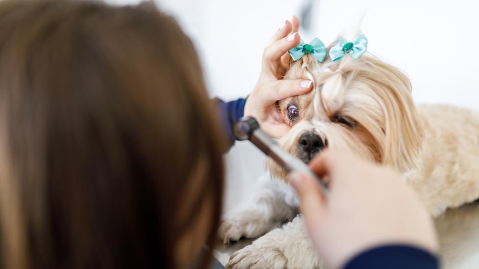
Anisocoria is diagnosed through an ophthalmic examination. Your veterinarian will use an instrument called an ophthalmoscope to examine the interior of your dog’s eyes. They will likely shine a bright light into each eye to evaluate the normal pupillary light response, to determine which pupil is normal and which is abnormal, and to see how well they constrict and dilate. They will do a full physical exam before deciding if more tests are necessary.
Your veterinarian will likely also perform several tests on your dog’s eyes. They may stain the cornea with fluorescein dye to detect ulcers, assess the pressure of the eyes, and check tear production. Depending on what they suspect the underlying cause to be, they may recommend blood work or imaging, especially if an underlying systemic disease is suspected.
Treatment of Anisocoria in Dogs
The treatment approach depends on the underlying cause and can vary greatly, ranging from no recommended treatment to surgery.
Many conditions that can lead to anisocoria, such as corneal injuries or damage to sympathetic nerves, can be effectively treated. Some conditions, like uveitis and glaucoma, can only be managed long-term, but anisocoria may resolve with appropriate management of the underlying disease.
Other conditions may be untreatable, such as iris atrophy or specific cancers. For iris atrophy, no treatment is necessary, as this is considered a benign change. This contrasts with a brain tumor, where treatment may not be feasible if a decrease in quality of life is likely.
Your veterinarian will discuss recommended treatment options after establishing a diagnosis. Anisocoria is a sign of an underlying condition, not a disease itself. Therefore, the focus is not on treating anisocoria, but rather on addressing the problem that led to the difference in pupil size.
Recovery and Management of Anisocoria in Dogs
Expectations for recovery are dependent on the underlying cause of anisocoria. If it’s due to iris atrophy in a small-breed senior dog, the prognosis for a normal life is excellent. In cases of an injury affecting nerves or the eye resulting in anisocoria, recovery from the injury should lead to resolution of anisocoria.
Some dogs may require lifelong treatment, such as eye drops, if an underlying eye condition is diagnosed. For instance, glaucoma requires consistent use of eye drops and regular veterinary check-ups to monitor eye pressure for the remainder of a dog’s life.
If your dog loses vision because of their condition, the vision loss is usually permanent. While this is not a common outcome for most underlying causes of anisocoria, it can occur if conditions are left untreated for too long. For this reason, it’s important to consult with your veterinarian right away if you see your dog’s pupils aredifferent sizes.
Anisocoria in Dogs FAQS
What is temporary anisocoria in dogs?
Temporary anisocoria is when a dog’s pupils are different sizes for a short duration of time and then return to normal. This can happen if there is inflammation inside the eye or inflammation affecting the nerves involved in changing pupil size. If this inflammation subsides quickly and pupils return to their normal size, it is categorized as temporary anisocoria.
Can anisocoria in dogs go away on its own?
Anisocoria can occasionally resolve on its own; however, it’s important to consult with a veterinarian right away to rule out more serious underlying causes, such as trauma.
Why are my dog’s pupils different sizes?
The term for different-sized pupils in dogs is anisocoria. This condition can stem from various underlying factors, ranging from minor issues to potentially life-threatening ones. If your dog’s pupils are different sizes, it means that either inflammation, compression, or injury has occurred affecting the eye, the nerves connected to the eye, or even the brain.
Featured Image: iStock.com/Capuski
References
Help us make PetMD better
Was this article helpful?
