Atlantoaxial Instability in Dogs
What Is Atlantoaxial Instability in Dogs?
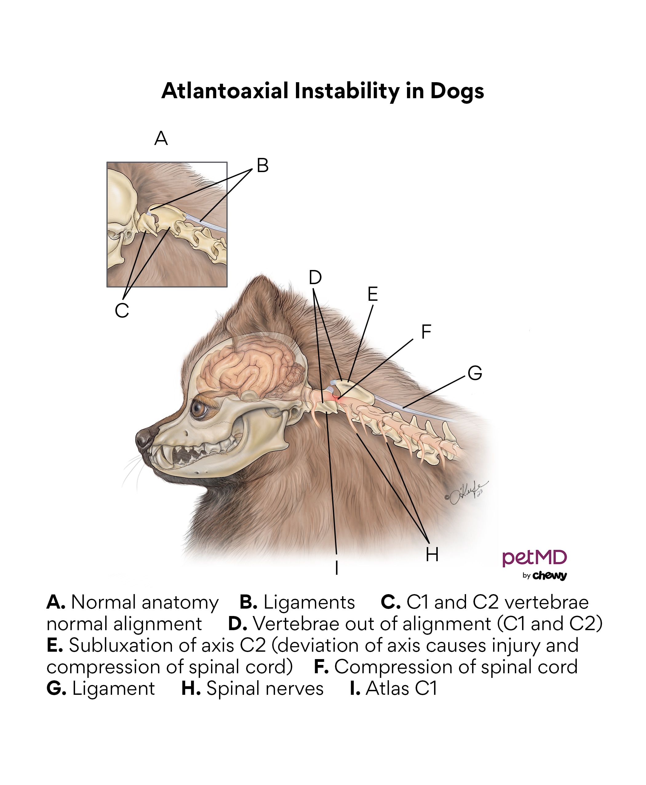
The atlantoaxial (AA) joint in dogs is located between the first and second neck vertebrae. The first vertebra, or atlas, and the second vertebra, or axis, are held together by ligaments (instead of a disc), which allows a dog to move their head from side to side.
While an uncommon condition, atlantoaxial instability, or AA instability (AAI), in dogs happens when the two bones are not aligned and cause friction by rubbing against the spinal cord and nerves, which results in severe neck pain and progressive neurologic symptoms.

Symptoms of Atlantoaxial Instability in Dogs
Dogs will typically have increasing neck pain—yelping or crying when touched—but they can also show the following symptoms:
-
Stiff neck
-
Low head carriage (head appears bowed)
-
Inability to stand or walk
-
Abnormal gait or ataxia (i.e., a drunken-like walk)
-
Difficulty or pain when eating or drinking
-
Rarely, apnea (lack of breathing), attributed to paralysis of the diaphragm
-
Death
It’s important to note that symptoms can be caused by injuries—even minor bumps—and symptoms can happen randomly or be seen all the time.
Causes of Atlantoaxial Instability in Dogs
AA instability is commonly due to a congenital birth defect, either in how the cervical bones (C1 and C2) fit together, the ligaments that connect them, or in the bones themselves. Additionally, trauma, such as being hit by a car, running into objects, or extreme rough play can cause the condition.
AA instability most often occurs in younger smaller-breed dogs such as Chihuahuas, Pekingese, Pomeranians, Toy Poodles, and Yorkshire Terriers, but the condition has also been seen in larger breeds like Rottweilers and Doberman Pinschers.
How Veterinarians Diagnose Atlantoaxial Instability in Dogs
In addition to a physical exam, where the dog will often exhibit pain in their neck and decreased range of motion, bloodwork and urinalysis screenings can be helpful in eliminating other causes and aid in further examination. A full neurologic exam will be performed to test reflexes and cranial nerves, and X-rays will most likely be recommended.
Referral to a veterinary neurologist may also be recommended to perform advanced imaging procedures such as CT, MRI, and even a CSF (cerebrospinal fluid) tap to help determine the extent of spinal cord injury.
Treatment of Atlantoaxial Instability in Dogs
For some dogs with minor discomfort or instability, conservative management with strict crate rest for multiple weeks, along with pain medications (such as gabapentin, tramadol, or other opioids, NSAIDs, or steroids) and a neck brace may be an option.
Surgery is, by far, the best option for dogs suffering from this condition, and even though it can be risky given the close proximity to the brain and spinal cord, it does provide the best chance of survival and long-term quality of life. The goal of the surgery is to minimize, if not eliminate, pain and to stabilize the joint, often through screws and bone cement. Dogs that have pre-existing medical conditions are not good candidates for anesthesia.
Without surgery, the prognosis for AAI is guarded, and symptoms can persist and progress to the point where quality of life is affected.
Recovery and Management of Atlantoaxial Instability in Dogs
For dogs being managed with medications only, it is critical that they be protected from any type of trauma, even minor bumps and scratches, as clinical signs can reoccur and have devastating consequences. Even with the utmost care and consideration, signs can recur and become progressive, as mentioned above.
Dogs that have undergone successful surgery can go on to live a functional and normal life, although refraining from high-impact activities or rough play is still advised to prevent further trauma or failure of the surgery.
Dogs that have neurologic signs or excessive dysfunction at the time of surgery have a much more guarded prognosis and may still suffer with neurologic problems afterward. The recovery period after surgery is often many weeks, with follow-up visits to the veterinarian for repeat X-rays to ensure proper healing and function. After surgery, dogs should be reintroduced to exercise gradually. Raised food and water bowls are helpful to minimize the amount of neck movement.
References
Help us make PetMD better
Was this article helpful?
