Liver and Gallbladder Cancer in Dogs
What is Liver and Gallbladder Cancer in Dogs?
Like many organs, the liver and gallbladder are susceptible to cancer. While a dog can have either liver cancer or gallbladder cancer, the unique location and function of these organs means that many dogs have cancer affecting both organs at the same time.
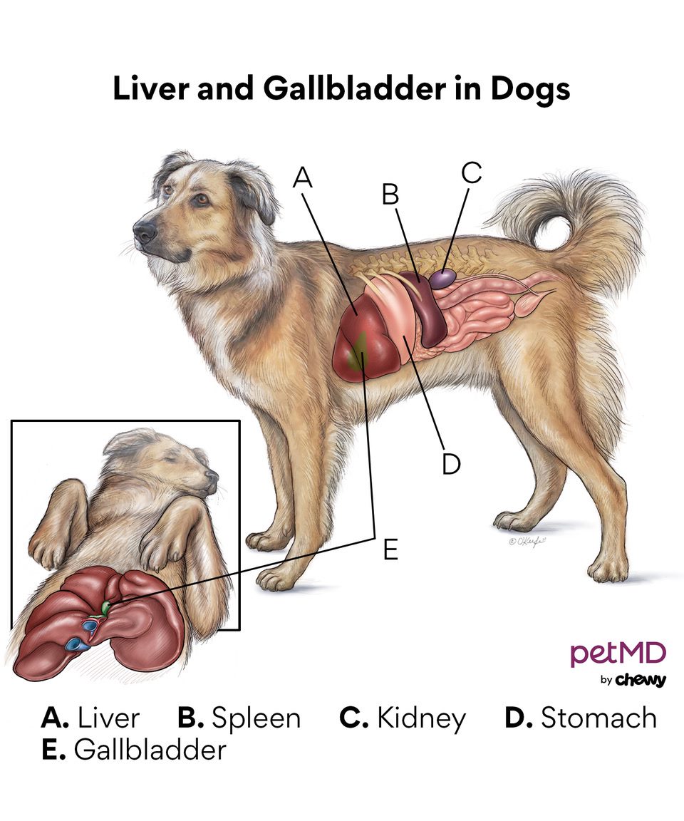
Dog livers have six lobes, with the gallbladder nestled between them. The liver and gallbladder sit in the abdominal cavity under the diaphragm and next to the stomach and pancreas.
Veterinarians class these types of tumors as:
-
Benign (non-cancerous)
-
Malignant (cancerous)
-
Primary (originated in the liver)
-
Secondary (spread from another tumor somewhere else)
-
Focal (located in one area)
-
Diffuse (spread throughout the entire organ)
Benign liver tumors are non-cancerous but are easy to confuse with malignant tumors. When veterinarians find any tumor in the liver, they may recommend additional tests to rule out more sinister diseases. Benign tumors do not spread to other parts of the body and do not pose a risk of metastasis. Many benign tumors may still be removed and dogs can have a good prognosis, if caught early. Benign tumors and lesions can be focal (located in one area or lobe) or diffuse and spread throughout multiple lobes. Examples of benign liver and gallbladder tumors include:
-
Hepatocellular adenoma
-
Hepatoma
-
Nodular hyperplasia
-
Bile duct adenoma
-
Hemangioma
Malignant Liver Tumors
While the prognosis for benign tumors is typically good if discovered early, the same is not true for malignant, cancerous tumors. The outcome for malignant tumors varies, based on how aggressive the cancer is in the dog. Malignant tumors can be primary, meaning it is a solitary cancer that may eventually spread to other sites. It may also be diagnosed as secondary which is metastasis from a distant tumor that has spread to the liver and gallbladder through the lymphatic system, blood vessels or direct invasion. Examples of malignant primary liver and gallbladder tumors include:
-
Hepatocellular carcinoma (this is the most common primary malignant liver tumor)
-
Cholangiocellular carcinoma
-
Hemangiosarcoma
-
Histiocytic sarcoma
-
Fibrosarcoma
-
Hepatic carcinoid
-
Lymphoma
Secondary malignant liver tumors are more common than primary malignant liver tumors. Other types of primary
-
Lymphoma
-
Intestinal carcinoma
-
Renal carcinoma
-
Mast cell tumors
-
Hemangiosarcoma
-
Islet cell carcinoma
-
Exocrine pancreas carcinoma

Symptoms of Liver and Gallbladder Cancer in Dogs
Clinical signs of liver cancer in dogs can be difficult to assess, nonspecific or even absent. If your dog does show signs of illness, symptoms may include:
-
Lethargy
-
Decreased appetite
-
Vomiting
-
Diarrhea
-
Low blood sugar, which may look like weakness, poor coordination or dullness
-
Weight loss
-
Ascites (abnormal fluid in the abdomen)
-
Increased thirst
-
Increased urination
-
Enlarged liver
-
Abdominal pain
-
Jaundice or icterus (yellowing of the skin and mucous membranes)
-
Collapse and acute shock due to tumor rupture
Causes of Liver and Gallbladder Cancer in Dogs
The definitive causes of this type of cancer are largely unknown. Genetics may play a role. Female dogs may be more prone to bile duct
How Veterinarians Diagnose Liver and Gallbladder Cancer in Dogs
Veterinarians may feel the tumor or fluid during an abdominal exam. They may also see a yellowing of the mucous membranes. A definitive diagnosis of liver or gallbladder cancer requires additional diagnostics.
Blood Chemistry and Complete Blood Count Testing
A complete blood count of a dog with liver or gallbladder cancer may reveal a decreased red blood cell count and changes to the platelets. The blood chemistry may reveal changes secondary to liver and gallbladder cell damage, bile stasis and liver insufficiency. Commonly, veterinarians see an elevation of liver- and gallbladder-specific blood values. Severity of blood changes doesn’t necessarily correlate with severity of disease, in all cases.
Bile Acid Testing
Bile acid tests assess the functioning of the liver and the results may be abnormal in dogs with liver cancer. The tests compare the bile acid level before and after a fatty meal to determine how well the liver is functioning and processing bile acids. Dogs with impaired liver function cannot process bile acids normally, resulting in high test results.
Coagulation Testing
The liver plays an active role in coagulation or clotting of the blood. Changes in liver function can directly impact the dog’s ability to clot normally.
Alpha-fetoprotein is a protein produced by young, regenerating or malignant liver cells. It is used as a marker of severe liver disease but doesn’t provide a definitive diagnosis of a specific type of cancer.
X-rays
Radiographs are useful to determine the overall size, shape and placement of the liver in reference to other abdominal organs. X-rays may allow visualization of obvious masses in the liver and other organs as well as abnormalities like calcification of abdominal contents. Veterinarians will also want to take X-rays of the lungs to look for a cancerous spread.
Ultrasound
Ultrasonography allows a more detailed analysis of the internal structure of the liver and other abdominal organs. Veterinary radiologists evaluate the entire abdominal cavity for other tumors, abnormalities and fluids. Veterinarians will also use ultrasound as a guide when they obtain biopsies, which is one way to get a definitive diagnosis.
Advanced Imaging
MRI (Magnetic Resonance Imaging) and CT (Computerized Tomography) scans help determine the location, size and severity of cancers within the abdominal cavity. They can sometimes suggest a certain type of cancer based on certain features on the scans. Advanced imaging is helpful in staging for surgery to allow full visualization of the tumor and how it interacts with the normal structures in the abdomen.
Surgery
Veterinarians use surgery to remove tumors or take biopsies of a tumor that cannot be removed completely through surgery. Typically, a dog will already have had blood work, X-rays and ultrasound, at a minimum, before surgery.
Treatment of Liver and Gallbladder Cancer in Dogs
Surgery
Surgery is the treatment of choice in dogs with primary tumors of the liver or gallbladder
The liver has amazing regenerative properties, so a dog can have a significant portion of the liver removed and still retain or regain function.
Tumors localized to just one liver lobe are easier to remove than tumors affecting multiple lobes. Veterinarians can also fully remove the gallbladder if necessary. Often, the location and type of tumor requires removing both the gallbladder and parts of the liver.
Before surgery, a complete work-up (cancer staging) should be performed to look at overall health and cancer spread. This may include blood work, X-rays of the chest, and an ultrasound of the abdomen. Based on the findings of these tests, vets will determine if a patient is a good candidate for surgery.
Chemotherapy
Dogs may be candidates for chemotherapy after removal of a liver and gallbladder tumor, or in unresectable tumors. Chemotherapy may also be an option to treat more advanced malignant secondary tumors.
Medication
No medication will cure liver or gallbladder cancer. However, some supplements, like S-Adenosyl-Methionine (SAMe) and Silybin (milk thistle) can help support the liver in regaining and protecting its function.
Prognosis of Liver Cancer and Gallbladder Cancer in Dogs
Based on the diagnosis, a prognosis of liver and gallbladder cancer may vary. Benign tumors typically have a good prognosis. If the tumor was found coincidentally, and if the pet is not actively sick, dogs may have an excellent full-life expectancy.
Patients who have additional secondary conditions may have a more guarded prognosis. Malignant and secondary tumors carry a more guarded-to-grave prognosis, based on the type of malignancy. In general, large single cancers carry a better prognosis than cancer that spreads to multiple nodules throughout the liver. Surgical resection can be curative, and life expectancy may be a few years.
Mesenchymal and neuroendocrine tumors have a worse prognosis than hepatocellular, with life expectancy of a few weeks to months. Gallbladder cancer has a high metastatic (spread) rate and is, in general, more difficult to remove than liver tumors.
Not all tumors can be removed. When they are not removed, tumors may grow so large that they rupture. Liver tumors tend to be fragile and vascular. When they rupture, dogs typically display:
-
Lethargy
-
Collapse
-
Pale gums
-
Increased heart rate
It is essential to seek veterinary care immediately if your dog is displaying any of these signs.
Recovery and Management of Liver and Gallbladder Cancer in Dogs
Recovery and management of liver and gallbladder cancer in dogs varies based on the treatment performed and type of tumor. It commonly involves addressing all symptoms
After surgery, a dog will require normal post-operative care relative to incision monitoring and routine pain management. The dog will likely have an incision from the sternum down to the pelvis.
These patients should have decreased activity and incision monitoring for at least 10 to 14 days. Re-checks with surgeons and oncologists may be scheduled initially at two weeks and then every one to three months going forward, depending on the plan. Veterinarians will likely check bloodwork parameters, X-rays and ultrasounds at visits to monitor disease progression and quality of life.
Liver and Gallbladder Cancer in Dogs FAQs
How long do dogs live with liver cancer?
Depending on the type of cancer, some dogs can live years after diagnosis. However, in more severe cases, the survival rate is much lower.
Is a dog with liver cancer in pain?
Depending on the specific disease, dogs with liver cancer may feel lethargic, appear nauseous and have a tender abdomen.
How fast does liver cancer progress in dogs?
Depending on the type, liver cancer can progress in a matter of weeks or years.
References
Help us make PetMD better
Was this article helpful?
